Mary Rutherford Imaging
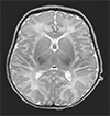
Magnetic resonance imaging (MRI) is now the imaging method of choice to provide detailed anatomical information about the fetal and neonatal brain, complimenting the screening role of ultrasound. MRI may be used to: –
- Confirm normal development
- Identify malformations of the brain
- Detect acquired injury to the brain
- Ascertain information on timing and aetiology of an abnormality
- Obtain a diagnosis and predict prognosis for the child
It is not however always easy to obtain good quality images in uncooperative, mobile patients. Poor quality examinations from motion artefact or poor signal to noise prevent full clinical interpretation and hamper the ability to predict the longer term outcome for the child. Progress has been made however in terms of image acquisition with practical solutions for preventing or compensating for motion.
In our increasingly litigious age the provision of detailed information on the developing brain , confirmation of normal anatomy and the visualisation and potentially timing of the onset of acquired lesions is required to inform health professionals and families.


